CT Oral Cases In FRCR 2B: How To Approach Cross-Sectional Imaging

Computed tomography (CT) forms a significant part of modern radiology practice and frequently appears in the FRCR 2B oral examination.

CT cases test a candidate’s ability to interpret cross-sectional anatomy, identify pathology, and explain findings clearly under examiner questioning.

Developing a consistent approach to CT interpretation helps candidates remain structured and confident during the viva.

Challenges of CT Oral Cases

CT images often contain large amounts of information. Candidates may feel overwhelmed when asked to interpret complex anatomy in a limited time.

Common challenges include:

  • Identifying subtle abnormalities
  • Describing anatomical relationships
  • Avoiding premature conclusions

A systematic interpretation strategy helps manage these challenges.

Start With Image Orientation

Begin by identifying:

  • Imaging modality (CT)
  • Plane (axial, coronal, sagittal)
  • Use of contrast

This demonstrates safe radiological practice and helps orient both you and the examiner.

Review Structures Systematically

When describing CT images, follow a consistent pattern. For example, in abdominal CT cases:

Solid Organs
Review the liver, spleen, pancreas, kidneys, and adrenal glands.

Bowel
Assess bowel loops for dilatation, obstruction, or inflammatory changes.

Vessels
Evaluate major vessels for thrombosis, aneurysm, or abnormal enhancement.

Other Findings
Look for lymphadenopathy, free fluid, or incidental findings.

This structured approach ensures important abnormalities are not overlooked.

Avoid Jumping to Conclusions

A common mistake in oral exams is offering a diagnosis too early.

Instead:

  1. Describe findings objectively
  2. Identify key abnormalities
  3. Suggest possible diagnoses

This sequence demonstrates logical reasoning.

Communicating Findings Clearly

When presenting CT findings verbally, clarity is essential.

For example:

“There is a dilated small bowel loop measuring approximately 3 cm with an abrupt transition point in the distal ileum, suggesting small bowel obstruction.”

Precise language improves communication and helps examiners follow your reasoning.

Practising CT Oral Cases

Cross-sectional imaging interpretation improves with exposure to multiple exam-style cases.

Structured case practice allows candidates to:

  • Improve anatomical orientation
  • Refine diagnostic reasoning
  • Develop confident verbal explanations

Explore our structured FRCR oral case.

Final Thoughts

CT oral cases test more than image interpretation. They assess a candidate’s ability to organise information, communicate clearly, and justify clinical reasoning.

Consistent case-based practice remains the most effective preparation strategy.

Author

Written by FRCR-qualified radiologists with experience in exam preparation and training.

Other Online Insights & Guides To Support FRCR 2B Oral Cases

Improving Verbal Communication For Frcr 2b Oral Exams

Learn how to improve verbal communication & structured explanations for the FRCR 2B oral exam.

How To Prepare Specifically For The Frcr 2b Oral Exam

Learn how to prepare effectively for the FRCR 2B oral exam. Improve structured reporting, viva confidence, diagnostic reasoning with practical strategies.

How To Justify Differential Diagnoses In Frcr Oral Exams

Learn how to prioritise & justify differential diagnoses clearly in FRCR 2B oral cases to maximise marks & demonstrate safe reasoning.

Nuclear Medicine Oral Cases For Frcr 2b

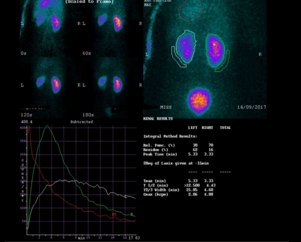
Discover how to approach nuclear medicine oral cases in the FRCR 2B exam, including radiotracer interpretation and structured reasoning.

Chest X Ray Oral Cases

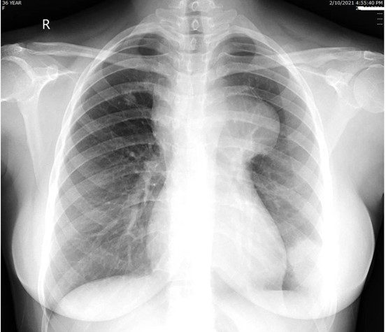
Learn how to approach chest X-ray oral cases in the FRCR 2B exam using a structured method that improves clarity, diagnostic reasoning, …

How To Structure Your Answers In Frcr Oral Cases

Discover a proven structure for answering FRCR 2B oral cases clearly & confidently. Improve your viva technique & exam performance.